
Horse health
The Horse’s Digestive System: How Gastric Ulcers Develop and What Owners Can Do About Them
How the horse’s forage-adapted digestive system works, why squamous and glandular gastric ulcers develop, the signs owners may notice, and how veterinary treatment and management can help.
By Debbie Irwin · 18 September 2026 · 7 min read
The horse’s digestive system is remarkably effective at extracting energy from fibrous plants, but it was designed around one fundamental assumption: the horse would spend much of the day moving and eating small amounts of forage. Modern management can be very different. Long periods without forage, concentrate meals, intensive exercise, travelling and restricted turnout can all alter the environment inside the horses stomach. For some horses, the result is gastric ulceration.
Equine Gastric ulcers are common enough that every horse owner should understand how the equine stomach works, why ulcers develop and importantly, why preventing them involves much more than simply reaching for an “ulcer supplement”.
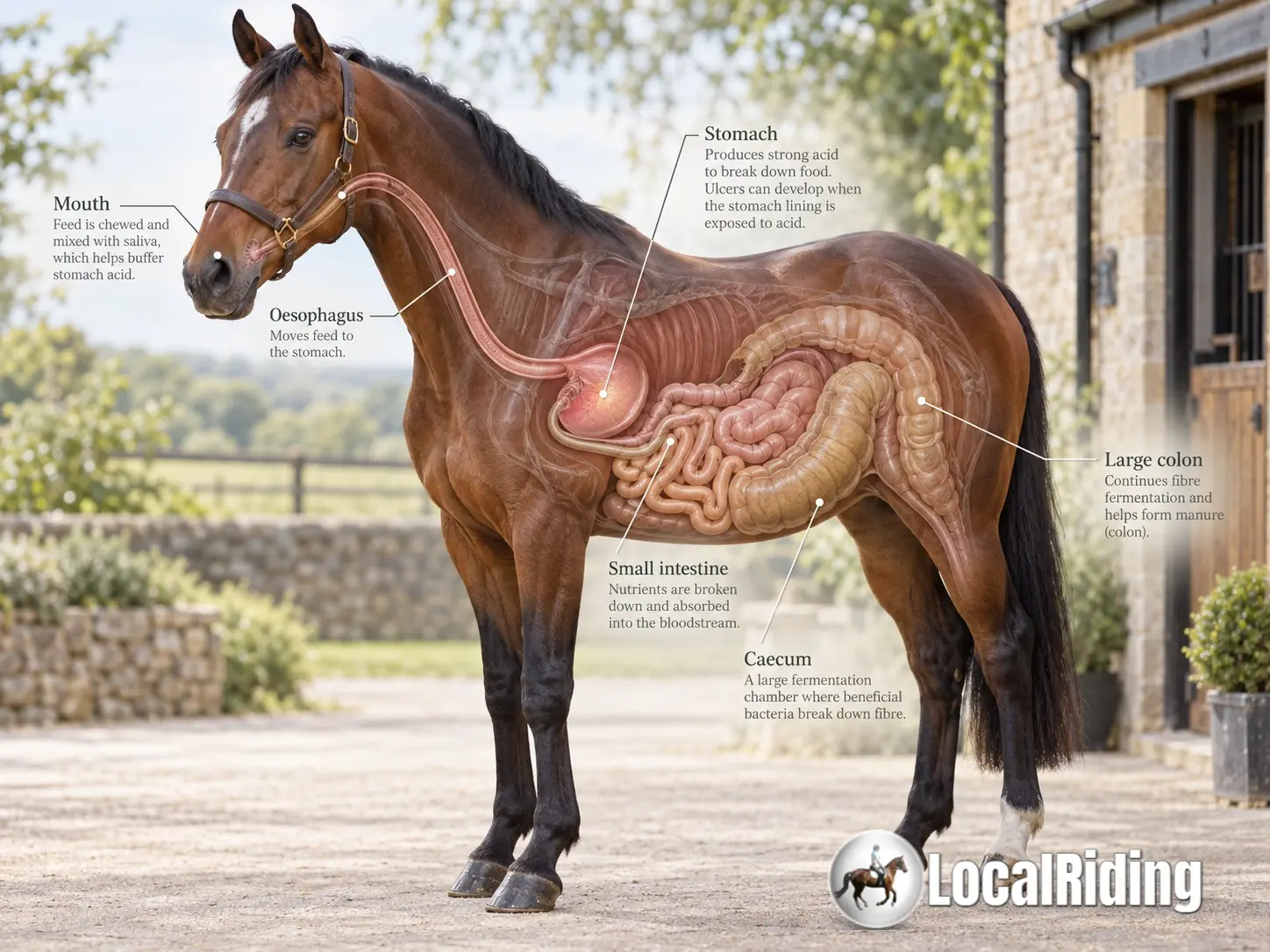 Labelled illustration of the horse digestive system, showing the mouth, oesophagus, stomach, small intestine, caecum and large colon
The horse’s digestive system is adapted for frequent forage intake and hindgut fermentation.
A digestive system built for forage
The horse is a non-ruminant herbivore and hindgut fermenter. Unlike cattle and sheep, it has a single stomach. Unlike people, however, a substantial proportion of the horse's energy can be obtained by microbial fermentation of fibre in the caecum and large colon.
The equine digestive journey begins before the feed reaches the stomach.
The Equine Mouth and Saliva
Horses are designed to chew for many hours each day. Grinding forage reduces particle size, but chewing also performs another extremely important function: it stimulates the production of saliva.
Saliva lubricates the feed and it contains bicarbonate and other substances that help buffer acidity within the stomach.
This creates an important relationship:
More time chewing forage → more saliva → greater buffering of gastric acid.
A horse consuming a forage-rich diet therefore spends considerably more time chewing, than the horse consuming concentrated feed to produce the same amount of energy.
Oesophagus
Once swallowed, feed travels down the oesophagus into the horses stomach.
The anatomy of the junction between the oesophagus and the stomach makes vomiting extremely difficult for horses. Food is essentially intended to travel in only one direction through the digestive tract.
The equine stomach — is surprisingly small
For an animal weighing perhaps 500–600 kg, the horse has a remarkably small stomach, with a capacity of only around 8–10 litres. It is designed to receive a fairly continuous stream of relatively small meals rather than one or two large feeds.
This becomes particularly important when we consider gastric ulcers.
The small intestine
From the stomach, partially digested food enters the small intestine: the duodenum, jejunum and ileum.
The jejunum alone is approximately 19.5 metres long. Enzymatic digestion takes place here and many nutrients, including amino acids, fats, vitamins, minerals and the products of carbohydrate digestion, are absorbed.
The hindgut
What remains then enters the caecum and large colon.
The caecum is effectively a large fermentation chamber holding approximately 27–30 litres. It contains a complex microbial population capable of breaking down plant fibre that the horse's own digestive enzymes cannot digest.
The resulting volatile fatty acids are absorbed and provide the horse with an important source of energy.
This is why forage is not simply something added to a horse's ration to keep it occupied. Forage fibre is fundamental to the way the equine digestive system evolved to function.
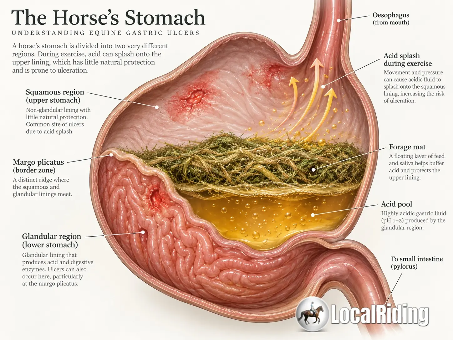 Labelled equine stomach anatomy showing the squamous region, glandular region and margo plicatus
The upper squamous and lower glandular regions have different protection from gastric acid.
The horse's unusual stomach
Understanding ulcers requires looking more closely at the stomach itself.
The equine stomach is divided into two distinctly different regions.
The squamous region
The upper portion is lined by squamous mucosa. Its surface is rather more like the lining of the oesophagus than the acid-producing portion of the stomach.
Crucially, it does not possess the same sophisticated acid-protection mechanisms as the lower stomach.
The glandular region
The lower portion is the glandular stomach.
This is where hydrochloric acid is produced. Because these tissues normally exist in an extremely acidic environment, they also possess protective mechanisms including mucus, bicarbonate, good mucosal blood flow and prostaglandin-dependent defences.
Separating the two areas is a clearly defined ridge known as the margo plicatus.
This anatomical divide explains why veterinary medicine now distinguishes two principal forms of gastric disease:
Equine Squamous Gastric Disease — ESGD and Equine Glandular Gastric Disease — EGGD.
Both fall within the broader term Equine Gastric Ulcer Syndrome, or EGUS, but they are not simply two versions of exactly the same disease. Their underlying mechanisms differ and this can affect both management and treatment. (Merck Veterinary Manual)
Horses produce stomach acid continuously
One of the most important facts for an owner to understand is that the horse does not produce gastric acid only when presented with a meal.
Acid secretion continues throughout the day.
That makes perfect evolutionary sense in an animal that would naturally graze for many hours.
When the horse is eating forage, several protective things happen simultaneously:
- Chewing produces saliva
- Saliva helps buffer gastric contents
- Forage remains within the stomach
- The fibrous contents help reduce exposure of the upper stomach to acidic fluid
Problems can arise when acid continues to be produced but the horse stops eating for prolonged periods. The stomach becomes emptier, saliva production decreases, acidic fluid remains, and the relatively vulnerable squamous lining can become exposed.
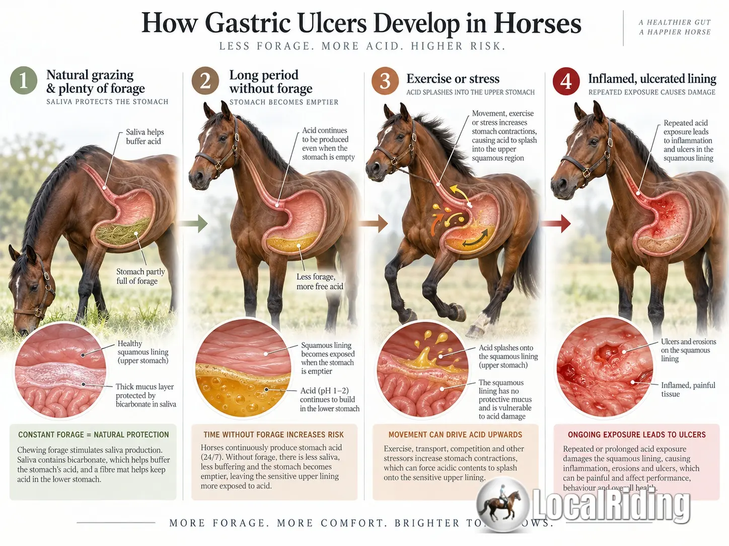 Four-stage illustration showing how reduced forage, continuous acid and exercise can contribute to equine gastric ulcers
How reduced forage and repeated acid exposure can progress from a healthy lining to squamous ulceration.
How a gastric ulcer actually forms
An ulcer is more than simply an irritated stomach. Repeated chemical injury first damages the surface of the stomach lining. Continued exposure can cause erosion and eventually create deeper lesions in the tissue.
Squamous ulcers: acid where acid should not be
ESGD is primarily an acid-exposure disease.
The glandular lower stomach is accustomed to an acidic environment. The squamous upper stomach is much less well protected. When acidic gastric contents repeatedly contact that upper lining, its cells can become damaged.
Several management practices make this more likely:
- An empty stomach: Periods without forage reduce chewing and saliva production while gastric acid secretion continues. Current veterinary nutritional guidance recommends avoiding periods of more than approximately 4 hours without forage.
- Exercise and the "acid splash" effect: As abdominal pressure rises during faster or more strenuous exercise, the contents of the lower stomach can be pushed upwards onto the unprotected squamous lining.
- High-concentrate diets: Large grain or concentrate meals create another problem by changing gastric fermentation, increasing acidic by-products, and requiring far less chewing.
Glandular ulcers are different
It is tempting to assume that all stomach ulcers simply result from excessive acidity. That is too simplistic.
The glandular stomach is supposed to contain acid. Its problem arises when the mechanisms protecting its own tissues become compromised.
With EGGD, attention turns to failure of the mucosal defence system:
- Disturbance of the mucus-bicarbonate barrier
- Reduced mucosal blood flow
- Altered prostaglandin activity
- Local inflammatory responses and impaired cellular repair
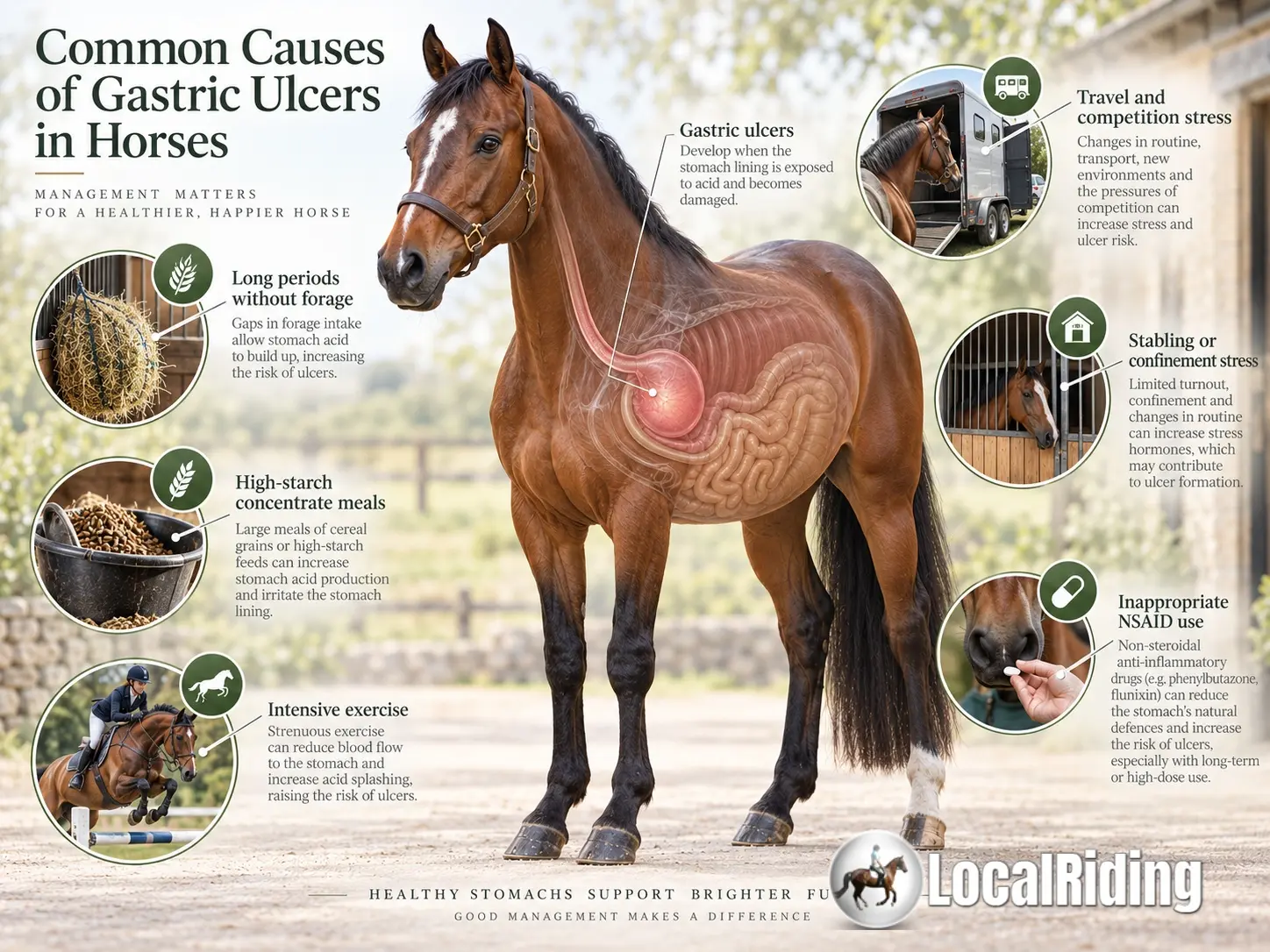 Illustrated guide to common equine gastric-ulcer risk factors
Several ordinary feeding, exercise and management factors may combine to increase risk.
What puts a horse at greater risk?
There is rarely one single culprit. Ulcers frequently arise when several relatively ordinary management factors occur together:
- Restricted forage: A horse that finishes its hay at midnight and is not fed again until 7am spends hours producing stomach acid with little buffer.
- Intensive work: Frequent strenuous exercise subjects the stomach to repeated acid splash.
- Large concentrate meals: High starch and sugar combined with limited forage create hostile conditions.
- Transport and competition: Travel combines restricted feeding, reduced movement, unfamiliar stabling, and physiological stress.
- Social stress & changes in routine: Herd displacement, box rest, weaning, and changing yards all impact feeding behaviour.
- Inappropriate NSAID use: Prolonged or excessive use of painkillers like phenylbutazone can interfere with protective prostaglandin mechanisms.
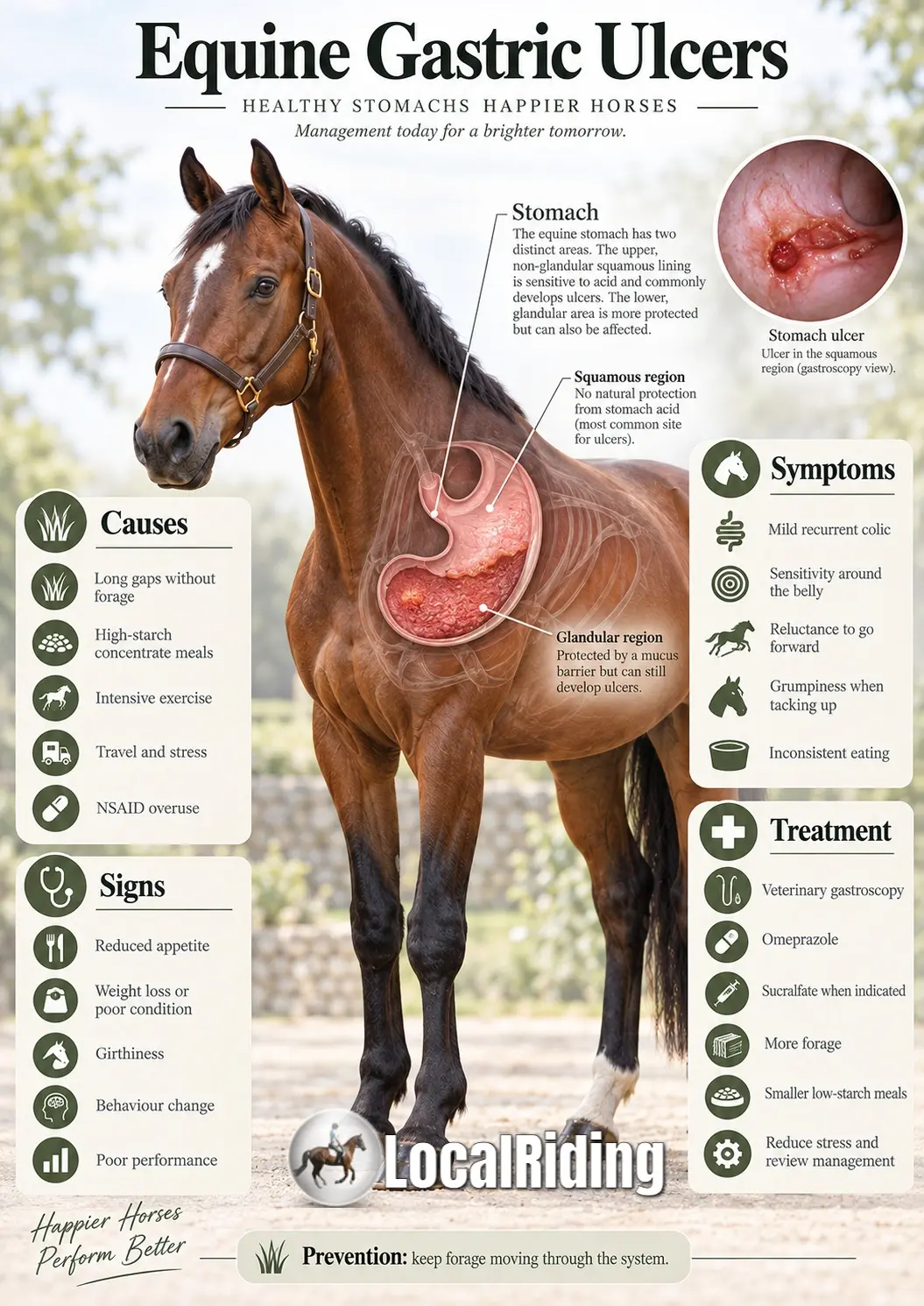 Equine gastric ulcer infographic summarising causes, signs, symptoms, diagnosis, treatment and prevention
Signs and symptoms
There is no single reliable outward sign of gastric ulcers. Some horses with substantial lesions show surprisingly little obvious discomfort, while others display behaviour or performance changes with relatively modest disease.
Common signs reported in veterinary references:
- Reduced or inconsistent appetite
- Poor body condition or mild weight loss
- Recurrent mild abdominal discomfort or colic
- Reduced performance and loss of impulsion
- Girthiness, tension, or resentment when tacked up
- Changes in attitude or temperament
Diagnosis: looking inside the stomach
The definitive method of diagnosing gastric ulcers is gastroscopy using a flexible endoscope passed into the stomach.
Gastroscopy tells the veterinarian which disease is actually present (ESGD vs. EGGD), allowing precise medical management.
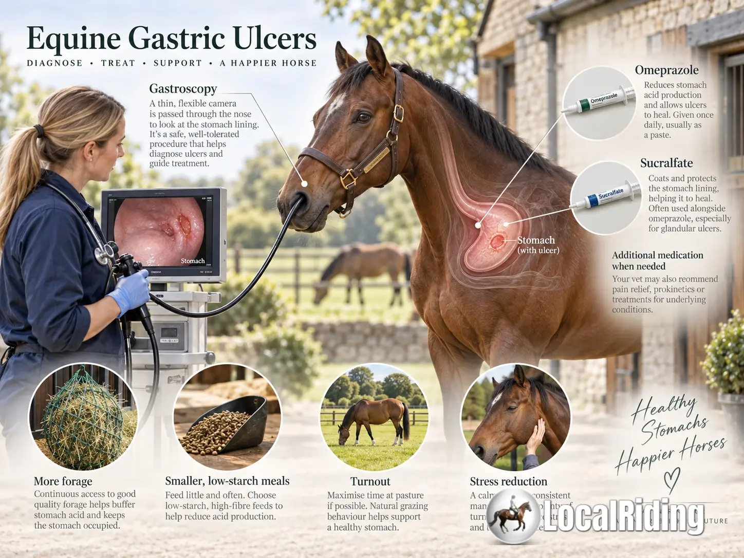 Veterinary gastroscopy and equine gastric-ulcer treatment and management overview
Treating gastric ulcers
Treatment combines veterinary medication with management changes:
- Omeprazole: The principal medication used for equine gastric ulceration. It suppresses hydrochloric-acid secretion, giving damaged squamous tissue an opportunity to heal.
- Mucosal protectants (sucralfate, misoprostol): Often required for glandular disease where acid suppression alone is insufficient.
Practical management principles
- Keep suitable forage moving through the system: Avoid periods exceeding 4 hours without forage. Use soaked hay, slow feeders, or small-holed nets for good doers.
- Don't exercise on an empty stomach: Feeding a small handful of lucerne/alfalfa chaff before riding provides a physical and acid-buffering mat.
- Reconsider starch before adding feed: Prioritise digestible fibre and oil over high-starch cereal mixes.
- Feed concentrates little and often: Split hard feed into multiple smaller meals.
- Preserve turnout and social stability: Freedom of movement and low-stress herd dynamics support healthy digestion.
This article is intended for general horse-owner education and is not a substitute for veterinary diagnosis or treatment. A horse showing persistent changes in appetite, behaviour, body condition, or performance should be assessed by a veterinary surgeon.
Debbie Irwin
Livery Yard Manager & Equestrian Professional
Debbie has run professional showing yards and mixed livery yards in Lincolnshire and Essex for over 30 years. She has competed and taught at various levels. She writes about practical horse health, yard life, livery and everyday horse health and management.